Cardionerds: A Cardiology Podcast

250. ACHD: Partial Anomalous Pulmonary Venous Return (PAPVR) with Dr. Ian Harris
Partial anomalous pulmonary venous return refers to anomalies in which one or more (but not all) of the pulmonary veins connects to a location other than the left atrium. This causes left to right shunting which may have hemodynamic and therefore clinical significance, warranting repair in some patients.
Join CardioNerds to learn about partial anomalous pulmonary venous return! Dr. Dan Ambinder (CardioNerds co-founder), Dr. Josh Saef (ACHD FIT at the University of Pennsylvania and ACHD Series co-chair), and Dr. Tripti Gupta (ACHD FIT at Vanderbilt University and episode lead) learn from Dr. Ian Harris (Director of the Adult Congenital Heart Disease program at University of California, San Francisco). Audio editing by CardioNerds Academy Intern, student doctor Shivani Reddy.
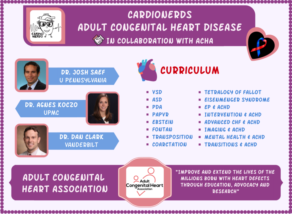
The CardioNerds Adult Congenital Heart Disease (ACHD) series provides a comprehensive curriculum to dive deep into the labyrinthine world of congenital heart disease with the aim of empowering every CardioNerd to help improve the lives of people living with congenital heart disease. This series is multi-institutional collaborative project made possible by contributions of stellar fellow leads and expert faculty from several programs, led by series co-chairs, Dr. Josh Saef, Dr. Agnes Koczo, and Dr. Dan Clark.
The CardioNerds Adult Congenital Heart Disease Series is developed in collaboration with the Adult Congenital Heart Association, The CHiP Network, and Heart University. See more
Disclosures: None
Pearls • Notes • References • Guest Profiles • Production Team

CardioNerds Adult Congenital Heart Disease Page
CardioNerds Episode Page
CardioNerds Academy
Cardionerds Healy Honor Roll
CardioNerds Journal Club
Subscribe to The Heartbeat Newsletter!
Check out CardioNerds SWAG!
Become a CardioNerds Patron!
Pearls – Partial Anomalous Pulmonary Venous Return (PAPVR)
- What is partial anomalous pulmonary venous return (PAPVR)?
- PAPVR refers to anomalies in which one or more (but not all) of the pulmonary veins connects to a location other than the left atrium. Often, this means one or more pulmonary veins empty into the right atrium or a systemic vein such as the superior vena cava or inferior vena cava. Physiologically, this produces a left-to-right shunt, allowing for already-oxygenated blood to recirculate into the lungs and result in excessive pulmonary blood flow.
- PAPVR refers to anomalies in which one or more (but not all) of the pulmonary veins connects to a location other than the left atrium. Often, this means one or more pulmonary veins empty into the right atrium or a systemic vein such as the superior vena cava or inferior vena cava. Physiologically, this produces a left-to-right shunt, allowing for already-oxygenated blood to recirculate into the lungs and result in excessive pulmonary blood flow.
- What are the clinical features of PAPVR?
- Diagnosis is usually incidental on a cross sectional imaging such as CTA or CMR.
- The most common associated lesion is an atrial-level defect.
- It is unusual for a single anomalous pulmonary venous connection of only 1 pulmonary lobe to result in significant shunting.
- Patients with a significant degree of left to right shunting may have right heart dilatation or symptoms of dyspnea on exertion.
- Diagnosis is usually incidental on a cross sectional imaging such as CTA or CMR.
- When are some strategies for managing patients with PAPVR?
- A surgical correction is recommended for patients with PAPVR when functional capacity is impaired and RV enlargement is present, there is a net left-to-right shunt sufficiently large to cause physiological sequelae (aka: ratio of pulmonary flow (Qp) to systemic flow (Qs) is > 1.5:1), PA systolic pressure is less than 50% systemic pressure and pulmonary venous resistance is less than one third of systemic venous resistance.
- Surgical repair involves intracaval baffling of the left atrium (Warden procedure) or direct reimplantation of the anomalous pulmonary vein into the left atrium.
- Pregnancy is well tolerated in patients with repaired PAPVR. In patients with unrepaired lesion who may have right sided heart dilatation and/or pulmonary hypertension, preconception evaluation and counseling should address how pregnancy may affect mother’s and fetus’s health.
- Antibiotic prophylaxis for infective endocarditis is typically not needed unless patients are less than 6 months from recent surgery, have residual defect at the patch margin or prior history of infective endocarditis.
- A surgical correction is recommended for patients with PAPVR when functional capacity is impaired and RV enlargement is present, there is a net left-to-right shunt sufficiently large to cause physiological sequelae (aka: ratio of pulmonary flow (Qp) to systemic flow (Qs) is > 1.5:1), PA systolic pressure is less than 50% systemic pressure and pulmonary venous resistance is less than one third of systemic venous resistance.
Show notes – Partial Anomalous Pulmonary Venous Return (PAPVR)
Notes (drafted by Dr. Tripti Gupta):
1. What is partial anomalous pulmonary venous return?
- Anatomically, partial anomalous pulmonary venous return refers to anomalies in which one or more (but not all) of the pulmonary veins connects to a location other than the left atrium. Often, this means one or more pulmonary veins empty into the right atrium or a systemic vein such as the superior vena cava (SVC) or inferior vena cava (IVC).
- Physiologically, this produces a left-to-right shunt, allowing for already-oxygenated blood to recirculate into the lungs and result in excessive pulmonary blood flow.
- If all pulmonary veins from both lungs drain to an anomalous site or in an abnormal fashion, then it is identified as a total anomalous pulmonary venous return (TAPVR). Patients with TAPVR often require surgical intervention in childhood.
- A bit of a nuance in terminology – partial anomalous pulmonary venous return (PAPVR) vs. partial anomalous pulmonary venous connection (PAPVC), requires some explanation. The suffix “return” refers to vessels returning to a chamber (ex: pulmonary vein returns to morphological left atrium after blood functionally mixes with systemic venous return or is redirected via an atrial septal defect) vs. “connection” implies abnormal anatomic attachments.
- Physiologically, this produces a left-to-right shunt, allowing for already-oxygenated blood to recirculate into the lungs and result in excessive pulmonary blood flow.
2. How does this happen? What is the embryological explanation for PAPVR?
- We know that the pulmonary veins originate from the posterior aspect of the left atrium. Meanwhile, the lung buds that arise from the lung parenchyma canalize as a vessel and gradually connect to the developing pulmonary veins.
- Some theories say that the lung buds are initially enmeshed in the splanchnic plexus which drains into the cardinal and umbilical vitelline veins (systemic venous system). By week 4 of gestation, the pulmonary veins from the left atrium connects with the superior portion of the splanchnic plexus to form the pulmonary plexus and ultimately loses its connection with the splanchnic plexus.
- The pulmonary vein is then supposed to divide into 4 branches, 2 on right and 2 on left, each with an orifice at the left atrium. Failure of one or more of the pulmonary veins to separate from the systemic venous systemic results in PAPVC/TAPVC.
3. What are some major clinical findings in PAPVR?
- PAPVR is typically an incidental diagnosis on CT or MRI in asymptomatic patients when these scans are done for another reason. Many patients with PAPVR may remain asymptomatic throughout childhood and adult life.
- Physiological changes may depend on degree of left to right shunt, number of veins involved, their sites of connection and associated lesions.
- 80% of anomalous connections are of the right sided pulmonary veins and 20% affect the left sided pulmonary veins. The most common variants include:
- Right upper pulmonary vein or right middle pulmonary vein to SVC, azygos vein, or right atrium. This variant is the most common and can be often associated with a sinus venosus defect.
- Right pulmonary veins to IVC, usually via a single trunk draining caudally and connecting to the IVC near the diaphragm. This variant is sometimes known as Scimitar syndrome. When you look at the descending trunk connecting the right venous return to the right atrium on x-ray or fluoroscopy, it has a crescent-like shape, like a Turkish sword from the Ottoman Empire or a scimitar, hence the name Scimitar syndrome.
- Left pulmonary vein(s) to the innominate vein via a vertical vein.
- Left pulmonary veins to the coronary sinus.
- Right upper pulmonary vein or right middle pulmonary vein to SVC, azygos vein, or right atrium. This variant is the most common and can be often associated with a sinus venosus defect.
- If more that 50% of a person’s pulmonary venous return drains anomalously to the right side of the heart, there may be right heart enlargement and presentation of symptoms such as dyspnea on exertion earlier in life.
- Physical exam findings may include prominent right ventricular impulse, a systolic ejection murmur at the left upper sternal border, split S2, and possibly a mid-diastolic rumble. In the absence of an ASD, these findings may not be obvious.
- On ECG, a RBBB morphology, RAD or first-degree heart block is associated with right ventricular volume enlargement.
- On echocardiogram, RV enlargement without left heart dysfunction should raise suspicion for anomalous pulmonary venous connection. Other hints can include the presence of a sinus venosus ASD, secundum ASD or RV enlargement that is significantly large for a small ASD/PFO. While left sided pulmonary veins can be visualized on the suprasternal view of transthoracic echocardiogram, right sided veins are more challenging on TTE. A TEE can be used to identify the site and drainage of pulmonary veins.
- A right heart catheterization is useful to identify the presence and etiology of pulmonary hypertension and quantify flows in pulmonary and systemic system and presence of a shunt. Selective angiography of the right and left pulmonary arteries can confirm the presence and course of pulmonary veins on levophase.
- A gated cardiac CTA or CMR is helpful and recommended for definitive diagnosis. A CTA offers higher spatial resolution than a CMR at the cost of radiation and iodinated contrast exposure. A CMR offers high resolution for defining vascular anatomy, quantifying chamber dimensions, estimate shunt burden and degree of stenoses using flow quantification techniques. In addition, respiratory-gated 3D whole heart imaging or MRA can be used for multiplanar reconstruction and aid in perioperative planning.
- Patients with TAPVR present with cyanosis at birth and need urgent surgical correction.
4. What conditions are associated with PAPVR?
- 80% of patients with PAPVR lesion may have an associated atrial level defect. In particular, a superior sinus venosus defect is frequently associated with right sided anomalous pulmonary venous connections.
- Other associated cardiac lesions include conotruncal abnormalities such as Tetralogy of Fallot or double outlet right ventricle, ventricular septal defects, and valvular abnormalities such as pulmonary stenosis, mitral or aortic stenosis or atresia and aortic arch anomalies.
- Anomalous pulmonary venous connections can also be seen in patients with heterotaxy syndrome, where mispositioned organs such as the heart, lungs, stomach, intestines, and liver may be in nonstandard locations within the chest and abdomen.
5. What are some main considerations for surgical repair for PAPVR?
- Cross sectional imaging such as CTA or CMR may be helpful to identify pulmonary venous connections and other extracardiac vascular anatomy.
- It is unusual for a single anomalous pulmonary venous connection of only 1 pulmonary lobe to result in a sufficient volume load to justify surgical repair. However, if a patient has symptoms referable to the shunt, there is >1 anomalous vein, and a moderate or large left-to-right shunt, then surgical repair is associated with a reduction in RV size and PA pressure. Pulmonary hypertension is a risk for adverse outcomes with surgery.
- A hemodynamic assessment with may help identify pressures, saturations, and degree of shunting.
- A surgical correction is recommended for patients with PAPVR when functional capacity is impaired and RV enlargement is present, there is a net left-to-right shunt sufficiently large to cause physiological sequelae (aka: ratio of pulmonary flow (Qp) to systemic flow (Qs) is > 1.5:1), PA systolic pressure is less than 50% systemic pressure and pulmonary venous resistance is less than one third of systemic venous resistance.
- Surgery can involve intracaval baffling of the left atrium (warden procedure) or direct reimplantation of the anomalous pulmonary vein directly into the left atrium.
- Repair of PAPVR may be considered at the time of closure of sinus venosus or other ASD.
- Transcatheter therapies are an area of ongoing innovation.
References – Partial Anomalous Pulmonary Venous Return (PAPVR)
- Gatzoullis MA, Webb GD, Daubeney PEF. Chapter 37: Partial Anomalous Pulmonary Venous Connections and Scimitar Syndrome In: Diagnosis and Management of Adult Congenital Heart Disease. 3rd ed. Elsevier Health Sciences; 2017: 354-361
- Kao CC, Hsieh CC, Cheng PJ et al. Total Anomalous Pulmonary Venous Connection: From Embryology to a Prenatal Ultrasound Diagnostic Update. J Med Ultrasound. Sep 2017; 25 (3): 130-137
- Pendela VS, Tan BEX, Chowdhury M, Chow M. Partial Anomalous Pulmonary Venous Return Presenting in Adults: A Case Series with Review of Literature. Cureus. 2020 Jun 1; 12 (6): e8388
- Stout KK, Daniels CJ, Aboulhosn JA, et al. 2018 AHA/ACC Guideline for the Management of Adults With Congenital Heart Disease: A Report of the American College of Cardiology/American Heart Association Task Force on Clinical Practice Guidelines. Circulation. Apr 2 2019;139(14):e698-e800.
Meet Our Collaborators!
Adult Congenital Heart Association
Founded in 1998, the Adult Congenital Heart Association is an organization begun by and dedicated to supporting individuals and families living with congenital heart disease and advancing the care and treatment available to our community. Our mission is to empower the congenital heart disease community by advancing access to resources and specialized care that improve patient-centered outcomes. Visit their website (https://www.achaheart.org/) for information on their patient advocacy efforts, educational material, and membership for patients and providers

CHiP Network
The CHiP network is a non-profit organization aiming to connect congenital heart professionals around the world. Visit their website (thechipnetwork.org) and become a member to access free high-quality educational material, upcoming news and events, and the fantastic monthly Journal Watch, keeping you up to date with congenital scientific releases. Visit their website (https://thechipnetwork.org/) for more information.

Heart University
Heart University aims to be “the go-to online resource” for e-learning in CHD and paediatric-acquired heart disease. It is a carefully curated open access library of educational material for all providers of care to children and adults with CHD or children with acquired heart disease, whether a trainee or a practicing provider. The site provides free content to a global audience in two broad domains: 1. A comprehensive curriculum of training modules and associated testing for trainees. 2. A curated library of conference and grand rounds recordings for continuing medical education. Learn more at www.heartuniversity.org/

CardioNerds Adult Congenital Heart Disease Production Team


 Amit Goyal, MD
 Daniel Ambinder, MD


 Visit Podcast Website
 RSS Podcast Feed
 Subscribe
 Add to MyCast