Cardionerds: A Cardiology Podcast

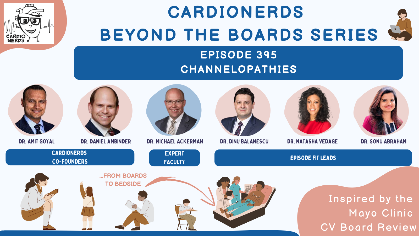
395. Beyond the Boards: Channelopathies with Dr. Michael Ackerman
Dr. Amit Goyal, along with episode chair Dr. Dinu Balanescu (Mayo Clinic, Rochester), and FIT leads Dr. Sonu Abraham (University of Kentucky) and Dr. Natasha Vedage (MGH), dive into the fascinating topic of channelopathies with Dr. Michael Ackerman, a genetic cardiologist and professor of medicine, pediatrics, and pharmacology at Mayo Clinic, Rochester, Minnesota. Using a case-based approach, they review the nuances of diagnosis and treatment of channelopathies, including Brugada syndrome, catecholaminergic polymorphic ventricular tachycardia (CPVT), and long QT syndrome. Dr. Sonu Abraham drafted show notes. Audio engineering for this episode was expertly handled by CardioNerds intern, Christiana Dangas.
The CardioNerds Beyond the Boards Series was inspired by the Mayo Clinic Cardiovascular Board Review Course and designed in collaboration with the course directors Dr. Amy Pollak, Dr. Jeffrey Geske, and Dr. Michael Cullen.
American Heart Association’s Scientific Sessions 2024
- As heard in this episode, the American Heart Association’s Scientific Sessions 2024 is coming up November 16-18 in Chicago, Illinois at McCormick Place Convention Center. Come a day early for Pre-Sessions Symposia, Early Career content, QCOR programming and the International Symposium on November 15. It’s a special year you won’t want to miss for the premier event for advancements in cardiovascular science and medicine as AHA celebrates its 100th birthday. Registration is now open, secure your spot here!
- When registering, use code NERDS and if you’re among the first 20 to sign up, you’ll receive a free 1-year AHA Professional Membership!
US Cardiology Review is now the official journal of CardioNerds! Submit your manuscript here.

CardioNerds Beyond the Boards Series
CardioNerds Episode Page
CardioNerds Academy
Cardionerds Healy Honor Roll
CardioNerds Journal Club
Subscribe to The Heartbeat Newsletter!
Check out CardioNerds SWAG!
Become a CardioNerds Patron!
Pearls and Quotes – Channelopathies
- One cannot equate the presence of type 1 Brugada ECG pattern to the diagnosis of Brugada syndrome. Clinical history, family history, and/or genetic testing results are required to make a definitive diagnosis.
- The loss-of-function variants in the SCN5A gene, which encodes for the α-subunit of the NaV1.5 sodium channel, is the only Brugada susceptibility gene with sufficient evidence supporting pathogenicity.
- Exertional syncope is an “alarm” symptom that demands a comprehensive evaluation with 4 diagnostic tests: ECG, echocardiography, exercise treadmill test, and Holter monitor. Think of catecholaminergic polymorphic ventricular tachycardia (CPVT) in a patient with exertional syncope and normal EKG!
- ICD therapy is never prescribed as monotherapy in patients with CPVT. Medical therapy with a combination of nadolol plus flecainide is the current standard of care.
- Long QT syndrome is one of the few clinical scenarios where genetic testing clearly guides management, particularly with respect to variability in beta-blocker responsiveness.
Notes – Channelopathies
1. What are the diagnostic criteria for Brugada syndrome (BrS)?
Three repolarization patterns are associated with Brugada syndrome in the right precordial leads (V1-V2):
- Type 1: Prominent coved ST-segment elevation displaying J-point amplitude or ST-segment elevation ≥2 mm, followed by a negative T wave.
- Type 2/3: Saddleback ST-segment configuration with variable levels of ST-segment elevation.
It is important to note that only a type 1 pattern is diagnostic for Brugada syndrome, whereas patients with type 2/3 patterns may benefit from further testing.
The Shanghai score acknowledges that relying solely on induced type 1 ECG changes has limitations. Therefore, one cannot equate the presence of a type 1 Brugada ECG pattern alone to the diagnosis of Brugada syndrome. The score suggests incorporating additional information—such as clinical history, family history, and/or genetic testing results—to achieve a definitive diagnosis.
2. What is the significance of genetic testing in Brugada syndrome?
There are 23 alleged Brugada syndrome susceptibility genes published with varying levels of evidence. However, only one gene mutation, the loss-of-function variants in the SCN5A gene encoding for the α-subunit of the NaV1.5 sodium channel, is considered to have sufficient evidence.
The overall yield of BrS genetic testing is 20%. The presence of PR prolongation (>200 ms) along with type I EKG pattern increases the yield to 40%. On the contrary, in the presence of a normal PR interval, the likelihood of SCN5A positivity drops to <10%.
3. How would you risk-stratify a patient with Brugada syndrome?
Serious arrhythmic events (SAE), including resuscitated cardiac arrest and sudden cardiac death, rarely represent the initial symptoms of Brugada syndrome. Thus, risk stratification is important.
Factors that increase risk include:
- Resuscitated cardiac arrest or history of cardiogenic syncope and the presence of a spontaneous type 1 ECG.
- Positive genetic test – certain SCN5A variants were independent predictors for SAE.
- Inducibility during programmed ventricular stimulation (EP study) using a double stimulation protocol (annualized risk is 1.5% per year for a positive study and 0.5% per year for a negative study).
4. What are the treatment options for Brugada syndrome?
- The only drug with therapeutic potential in BrS is quinidine. The antiarrhythmic effect is achieved by prolonging the effective refractory period via inhibition of Ito potassium channel.
- BrS plus a history of cardiac arrest, sustained VT, or syncope judged to be caused by ventricular arrhythmia would warrant an ICD.
- In those refractory to first-line therapies, RVOT epicardial ablation is now an additional therapeutic option.
5. What are the four diagnostic tests to be done in a patient who presents with an episode of exertional syncope?
Exertional syncope is a high-risk presentation that demands a comprehensive evaluation! This includes:
- EKG
- Echocardiogram
- Exercise treadmill test
- Holter monitor
Do not stop at an EKG and echo alone!
Think of catecholaminergic polymorphic ventricular tachycardia (CPVT) in a patient with exertional syncope and a normal EKG!
6. What are the features on the exercise treadmill test that increase the suspicion for CPVT?
Bidirectional VT is considered a hallmark of CPVT, with digoxin toxicity being the only real imitator. This finding is specific in the absence of digoxin but not sensitive.
During exercise testing in CPVT, as the patient’s heart rate rises with increasing workload, PVCs begin to appear, progressing to bigeminy, couplets, and, in some instances, bidirectional couplets. The ectopy typically vanishes within 30 seconds of the recovery phase. This pattern increases suspicion of CPVT and warrants a detailed family history and genetic testing.
7. What are the genetic underpinnings of CPVT?
Mutations in the ryanodine receptor (RyR2 gene) render calcium release channels leaky, leading to diastolic calcium overload. This ultimately triggers arrhythmias in CPVT.
8. What are therapeutic interventions for a patient with CPVT?
Medical therapy is the mainstay of treatment in CPVT. Drugs include non-selective beta-blockers like nadolol or propranolol. Standard of care currently includes a combination of nadolol plus flecainide. An ICD is indicated only in the case of an aborted cardiac arrest. ICD therapy is never prescribed as monotherapy in these patients.
9. How do we correctly measure the QTc?
The QT interval is measured from the beginning of the QRS complex to the end of the T wave. The end of the T wave is determined using the maximum slope intercept method, in which a tangent line is drawn through the maximum down slope of the T wave. The point at which this tangent line crosses the isoelectric line is the end of the T wave. The U wave is excluded.
- Best measured in leads II or V5.
- Quick eyeball test: if the QT interval is more than ½ the RR interval, the QTc will be >460 ms.
10. What are the three primary mutations implicated in Long QT syndrome?
- LQT1 (30-40% of cases)
- Mutation: loss of function in potassium channel gene KCNQ1
- ECG: broad-based T wave
- Trigger: activity, adrenaline, exercise
- BB responsiveness: +++ (nadolol or propranolol)
- LQT2 (second most common)
- Mutation: loss of function in potassium channel gene KCNH2
- ECG: notched T wave
- Trigger: auditory (alarm clock), post-partum
- BB responsiveness: ++
- LQT3
- Mutation: gain of function or leakiness of sodium channel SCN5A (note: BrS is due to loss of function in the same gene)
- ECG: normal T wave after prolonged isoelectric ST segment
- Trigger: none, but typically happens during rest
- BB responsiveness: + (propranolol); may consider combination therapy with mexiletine or mexiletine monotherapy.
References – Channelopathies
- Charles Antzelevitch, Gan-Xin Yan, Michael J. Ackerman, Arthur A.M. Wilde et al. J-Wave syndromes expert consensus conference report: Emerging concepts and gaps in knowledge, EP Europace, Volume 19, Issue 4, April 2017, Pages 665–694
- Krahn AD, Behr ER, Hamilton R, Probst V, Laksman Z, Han HC. Brugada Syndrome. JACC Clin Electrophysiol. 2022 Mar;8(3):386-405. PMID: 35331438.
- Chockalingam, P, Crotti, L, Girardengo, G. et al. Not All Beta-Blockers Are Equal in the Management of Long QT Syndrome Types 1 and 2: Higher Recurrence of Events Under Metoprolol. JACC. 2012 Nov, 60 (20) 2092–2099
- Priori SG, Wilde AA, Tracy C et al. HRS/EHRA/APHRS expert consensus statement on the diagnosis and management of patients with inherited primary arrhythmia syndromes: document endorsed by HRS, EHRA, and APHRS in May 2013 and by ACCF, AHA, PACES, and AEPC in June 2013. Heart Rhythm. 2013 Dec;10(12):1932-63. PMID: 24011539.
- Viskin S, Rosovski U, Zeltser D et al. Inaccurate electrocardiographic interpretation of long QT: the majority of physicians cannot recognize a long QT when they see one. Heart Rhythm. 2005 Jun;2(6):569-74. PMID: 15922261.
- Horner JM, Ackerman MJ. Ventricular ectopy during treadmill exercise stress testing in the evaluation of long QT syndrome. Heart Rhythm. 2008 Dec;5(12):1690. PMID: 19084807; PMCID: PMC3281579.


 Visit Podcast Website
 RSS Podcast Feed
 Subscribe
 Add to MyCast